Main Research Interests
My research interests focus on the morphology of the brain. I began my research career focusing on the endocranial (inner surface of the skull) morphology of modern humans, hoping to ground truth the relationship between the brain and the skull for paleoanthropological research. I have since switched my focus to modern brain morphology, specifically the morphology of the white matter of the brain. I am particularly interested in bringing shape analysis techniques used in anthropology and evolutionary biology to neuroanatomy research.
Doctoral Research
Current Projects
Brainlife.io: "We are developing an open, online platform to provide seamless access to cloud computing infrastructure, brain data, and data derivatives. This platform is meant to reach out beyond neuroscience, allowing also computer scientists, statisticians and engineers interested in brain data to use the data to develop and publish their methods."
As a core member of the brainlife.io developer team, I have developed several open-source applications for diffusion MRI data processing and shape analysis.
As a core member of the brainlife.io developer team, I have developed several open-source applications for diffusion MRI data processing and shape analysis.
|
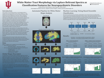
Shape analysis pipeline for studying the white matter tracts of the brain.
Featured in small article in the IU IT News & Events Presented at ShapeMI at MICCAI 2018 in Granada, Spain! Published online here. Author version PDF available here! Abstract: Diffusion-weighted magnetic resonance imaging (dMRI) allows for non-invasive, detailed examination of the white matter structures of the brain. White matter tract specific measures based on either the diffusion tensor model (e.g. FA, ADC, and MD) or tractography (e.g. volume, streamline count or density) are often compared between groups of subjects to localize differences within the white matter. Less commonly examined is the shape of the individual white matter tracts. In this paper, we propose to use the Laplace-Beltrami (LB) spectrum as a descriptor of the shape of white matter tracts. We provide an open, automated pipeline for the computation of the LB spectrum on segmented white matter tracts and demonstrate its efficacy through machine learning classification experiments. We show that the LB spectrum allows for distinguishing subjects diagnosed with bipolar disorder from age and sex matched healthy controls, with classification accuracy reaching 95%. We further demonstrate that the results cannot be explained by traditional measures, such as tract volume, streamline count, or mean and total length. The results indicate that there is valuable information in the anatomical shape of the human white matter tracts. |
|
|
First Year Project:
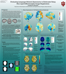
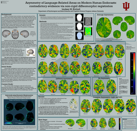
A more accurate measure of asymmetry of the modern human endocranium, with a focus on language related areas. Abstract: Topographical features of the endocranial surface are thought to correspond to the brain surface, meaning that changes in these features throughout hominid evolution can be used to infer evolutionary changes in brain structure. By examining changes in endocranial regions that directly overlay brain areas related to certain behaviors (i.e. language) we may be able to estimate the temporal development of those behaviors. There is an established leftward asymmetry of Broca’s area, one of the major language areas of the human brain, and previous qualitative research suggests that Broca’s cap (a region on endocasts near Broca’s area) is also larger on the left (Broadfield and Holloway, 2001). However, recent research using geometric morphometrics (GM) found a rightward trend for Broca’s cap in modern humans (Balzeau et al., 2014, Kitchell, 2015). This study investigated language-related asymmetry of the modern human endocast using non-rigid diffeomorphic image registration, a method typically used in neuroscience to register individual brains to a reference atlas. Although other quantitative methods exist to assess endocranial asymmetry, this method is distinct in that it is automated and does not require the manual placement of points (as in GM) or manual delineation of a region of interest (as in GIS) and thus can avoid potential bias. Results based on 100 endocasts of modern humans indicate that Broca’s cap is actually an average of 5% larger on the right in modern humans, contradictory to previous qualitative studies. However, a region directly above Broca’s area is an average of 3% larger on the left side, suggesting it may still be possible to see language related asymmetries on an endocast. These results suggest that we must reevaluate previous assumptions that Broca’s cap asymmetry is related to language lateralization and provide motivation for reexamining other qualitative claims about endocranial morphology using quantitative, relatively bias-free methods. |
|
Photogrammetry, 3D printing, and Archaeology:
As a member of El Proyecto Antropologico de Quiechapa, I created 3D models of archeological artifacts found during survey using various photogrammetry techniques. I prepared the 3d models of artifacts and landscapes (that were created via drone photography) for 3D printing and I 3D printed the archaeological artifacts as well as the landscape models using my personal 3D printer. I also explored the use of 3D printed landscape models while mapping sites during survey.
3D printing and 3D model creation (via CT scans, MRI scans, photogrammetry, laser scanning):
I am exploring the many ways 3D printing, 3D scanning, and 3D models of brain areas, bones, and artifacts can be beneficial to the fields of Neuroscience, Cognitive Science, Archaeology, Anthropology, and Paleoanthropology. There is a wealth of information freely available to be 3D printed, as well as many ways to create models from the data used in your own research. I post photos and information about many of the items I print on my instagram (https://www.instagram.com/paleoneuro/) and twitter (https://twitter.com/LindseyKitchell) accounts.
Master's Research
Asymmetry of the Modern Human Endocranium
Awarded Distinction and UCL Institute of Archaeology Master's Prize for Outstanding Dissertations
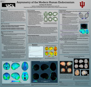
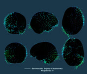
Abstract: Hominin brain evolution is a topic of great interest in paleoanthropology. Details of this evolutionary process are typically inferred from endocasts. However, very little research has been done to quantifiably establish the relationship between an endocast and its corresponding brain. This study investigates this relationship using asymmetry of the entire endocranial surface. As the modern human brain is structurally asymmetric, the results of this study allow for a direct quantitative comparison of this characteristic between the two surfaces. In addition, because important aspects of behavior, such as handedness and language processing, are organized asymmetrically in the brain, it is of great interest to be able to see these same asymmetries on the endocranial surface.
Using innovative geometric morphometric techniques, this study quantified the degree and direction of asymmetry of the entire endocranial surface in adult modern humans. Results indicate the well-known petalia pattern of asymmetry extends beyond the frontal lobe to include the right temporal and anterior parietal regions. In addition to an anterior-posterior and lateral asymmetry, the petalias also differ in superior-inferior distribution. A rightward asymmetry of Broca’s area was found, contradicting previous qualitative reports. A leftward asymmetry of the anterior cerebellum was also found and a rightward asymmetry of the temporal pole, as well as several other subtle asymmetries across the rest of the endocranial surface. The findings are compared with brain asymmetry research and the implications for brain evolution research are discussed.
View on Academia.edu
View instructions for the methods used at endocranialasymmetrymethods.blogspot.com

|
|
|
Undergraduate Research
Structural Asymmetry of the Human Brain Assesed via Non-rigid Diffeomorphic MRI Registration
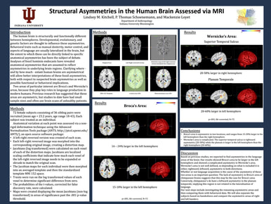
Abstract: The human brain is structurally and functionally different between hemispheres. Developmental, evolutionary, and genetic factors are thought to influence these asymmetries. Behavioral traits such as manual dexterity, motor control, and aspects of language are usually lateralized in the brain, but the extent to which these can be directly linked to specific anatomical asymmetries has been the subject of some debate. Analyses of fossil hominin endocasts have revealed anatomical asymmetries that are assumed to reflect asymmetries in underlying brain regions. Clarifying where – and by how much – extant human brains are asymmetrical will allow better interpretations of these fossil asymmetries, both with respect to suspected brain asymmetries as well as possible functional/behavioral implications. Two areas of particular interest are Broca’s and Wernicke’s areas, because they play key roles in language production in modern humans. Previous research has suggested that these areas are asymmetric, but studies to date have had small sample sizes and often use brain scans of unhealthy patients. To this end, we investigated the various left-right differences of the human brain through a voxel-based morphometric analysis of MRI scans of 66 healthy, female subjects. Left-right reversed versions of individual brains were mapped into their corresponding original versions, using non-rigid deformation methods. These mappings were then registered to a common atlas, and average degrees of left-right asymmetry were calculated for each voxel. Our results showed both Broca’s area and Wernicke’s area to have significant leftward asymmetry at the FDR corrected q-value of .01. Implications of this work for hominin evolution will be discussed.
Structural Asymmetries in the Human Brain Assessed via MRI
The Association Between Handedness and Structural Asymmetries of the Human Brain
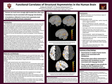
Abstract: The human brain is both structurally and functionally asymmetric. However there have been relatively few studies assessing both structural and functional asymmetries in the same subject population. One behavioral asymmetry of particular interest to human evolution is right-handedness, which is much more pronounced at the population level in humans than in other species. It has been postulated that this may be related to the evolution of tool manufacturing and use, and potentially also to language (which, like control of the right hand, is also lateralized to the left hemisphere). Analyses of fossil hominin endocasts have revealed anatomical asymmetries that are assumed to reflect asymmetries in underlying brain regions. Handedness has been shown to be associated with brain petalias in at least one study, but its association with other structural other areas of the brain have not been extensively investigated. Knowing the extent to which neuroanatomical asymmetries in different regions of the brain are associated with handedness will allow better assessment of handedness in fossil specimens. We report here the results of a study of 72 healthy, female subjects in which degree of handedness was correlated with neuroanatomical asymmetry at each point, assessed via non-rigid deformation (morphing) methods of their MRI scans. Areas of highest association between right-handedness and neuroanatomical asymmetry included: 1) the left motor cortex corresponding to control of the right hand, 2) left occipital pole and adjacent regions, 3) left parietal-occipital-temporal region (Wernicke’s area), 4) right middle temporal sulcus, and 5) left orbital frontal.
Functional Correlates of Structural Asymmetries in the Human Brain
Feature selection Strategies and Perceptual Expertise in Configuration Search Tasks
Abstract: Configurations of simple objects often play a role in real world visual search tasks. For example, diagnosing a dislocated joint in an x-ray or individualizing a fingerprint to a single person are both tasks in which the relative locations of features are more important than the identity of simple features. To study the nature of perceptual expertise in a search task in which configurations define the target, we collected eye-tracking data while participants played the video game Bejeweled 2. Participants excel at this task by attending to particular configurations of pieces and ignoring others. We analyzed the eye-tracking data at every 60ms interval. We defined a set of meaningful and misleading templates and determined how often experts and novices fixated on each. This feature induction reveals the nature of the strategies that underlie perceptual expertise in this domain and establishes this methodology as a means to uncover the feature set used by participants.
